26
Mar
How Intraoral Cameras Are Changing the Way Dentists Diagnose Problems
There was a time when a dental exam meant lying back, staring at the ceiling, and trusting that your dentist could see everything worth seeing with a handheld mirror and a light. That approach worked, but it had real limitations. At Seasons Dental in Burley, Idaho, intraoral cameras are now a routine part of how we examine teeth, and the difference in what we can catch, and how early we can catch it, is significant.
What an Intraoral Camera Actually Is
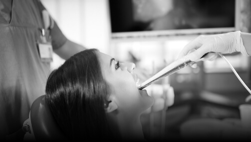
An intraoral camera is a small, pen-shaped device with a tiny camera at the tip. It fits comfortably inside the mouth and captures high-resolution images of the teeth, gums, and surrounding tissue in real time. Those images display on a screen immediately, giving both the dentist and the patient a detailed, magnified view of what is happening inside the mouth.
The camera itself is about the size of a large marker. It is lightweight, easy to maneuver, and produces images that are far more detailed than anything visible to the naked eye during a standard exam.
What It Changes About Diagnosis
Traditional visual exams rely entirely on what a dentist can see from a standing position with standard lighting. Small cracks, early decay forming between teeth, worn enamel, and failing margins around older restorations can all be easy to miss at that scale. An intraoral camera eliminates most of that uncertainty.
Hairline cracks in enamel are a good example. They rarely cause symptoms in early stages and are nearly invisible without magnification. Left undetected, a crack can deepen over time until it reaches the pulp or causes the tooth to fracture. Catching it early means the treatment is simple. Catching it late often means a crown or, in some cases, extraction.
The same applies to the margins of existing fillings and crowns. Over time, restorations can begin to separate slightly from the tooth structure beneath them, creating a gap where bacteria accumulate. That gap may be too small to feel and too subtle to see clearly without a magnified view, but an intraoral camera picks it up cleanly.
The Patient Experience Changes Too
One of the less obvious benefits of intraoral cameras is what they do for patient understanding. When a dentist describes a problem verbally, patients are largely taking it on faith. When a patient can see a cracked tooth or inflamed gum tissue on a screen in front of them, the conversation becomes entirely different.
People make better decisions about their own care when they understand what they are looking at. Seeing a fracture or early decay in real time removes the ambiguity from a treatment recommendation. It is not that patients distrust their dentist; it is that visual confirmation makes the situation concrete in a way that a description simply cannot.
How It Works Alongside Other Technology
Intraoral cameras do not replace X-rays or CBCT scanning. They serve a different function. X-rays reveal what is happening inside the tooth and below the gumline. A CBCT scan provides a three-dimensional picture of bone structure, root anatomy, and surrounding tissue. Intraoral cameras capture the surface in fine detail.
Used together, these tools give a much more complete diagnostic picture than any one of them provides alone. At Seasons Dental, combining intraoral imaging with digital X-rays means fewer surprises, earlier intervention, and treatment plans grounded in accurate information rather than estimation.
Early Detection Has a Direct Impact on Treatment Complexity
Dentistry follows a simple pattern: problems caught early are less expensive, less invasive, and easier to treat. A cavity identified when it is small gets a filling. The same cavity found a year later might need a crown. Found later still, it may require root canal therapy or extraction.
Intraoral cameras push detection earlier in that timeline. When a dentist can see a tooth clearly enough to identify the beginning of decay or a stress fracture before symptoms develop, patients get options they would not have had otherwise.
What to Expect During Your Visit
If you have not had a dental exam that includes intraoral imaging, the process is straightforward. The camera is guided around the mouth during the examination portion of your appointment. The images appear on a chairside screen, and your dentist will walk you through anything notable. It adds very little time to the visit and requires nothing on your part except keeping your mouth open.
For patients who have felt uncertain about dental recommendations in the past or who simply want a clearer picture of their oral health, intraoral cameras make the entire process more transparent. Seasons Dental uses this technology because better information leads to better care, and that benefits everyone in the chair.

Chad Bodily, DDS
Connect on LinkedInDr. Chad Bodily, DDS, is a compassionate dentist with strong ties to the Mini-Cassia community. After graduating from Minico High School and serving a church mission in Portugal, he earned a bachelor's degree in Biology from BYU-Idaho and a Doctorate of Dental Surgery from the University of Iowa. Dr. Chad partners with his brother, Dr. Ty, to provide patient-focused care, treating everyone like family. Committed to professional growth, he is licensed in sedation dentistry, ensuring a comfortable experience for his patients. Dr. Chad values building strong patient relationships and considers his family his greatest joy and accomplishment.

Ty Bodily, DMD
Connect on LinkedInDr. Ty Bodily, DMD, is a skilled dentist with deep roots in the Mini-Cassia area. A proud graduate of Minico High School and BYU-Idaho, he earned his Doctorate of Medical Dentistry from Nova Southeastern University in Florida. A highlight of his education was volunteering in Brazil, where he provided free dental care to underprivileged children. With post-graduate training from world-renowned experts in sedation, restorative, and cosmetic dentistry, he excels in reconstructing smiles, enhancing both health and self-esteem. Dr. Ty's passion for dentistry is matched only by his devotion to his family, whom he considers his greatest achievement and passion.